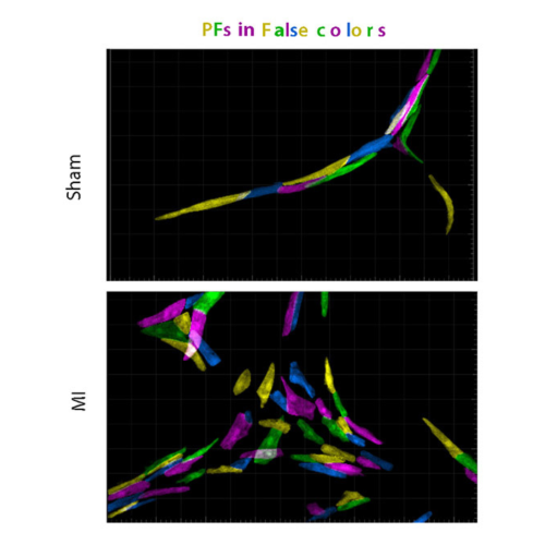
Myocardial infarction induces cardiomyocytes death and impaired cardiac function, which is followed by a high prevalence of life-threatening arrhythmia that is likely due to conduction defects. Unlike adult mammals, newborn mice can regenerate a functional heart after myocardial infarction. In this study published in Nature Cardiovascular Research, Lucie Boulgakoff from Kelly’s team, discovered that cardiac regeneration following neonatal infarction in mice was not so perfect concerning the electrical wiring of the heart that is known as the Purkinjer fiber network. Using fancy genetic tools, Lucie showed that after cardiac regeneration Purkinje fibers present a short and rectangular shape while they are thin and elongated in sham-operated hearts.
In summary, in newborn mice, after a neonatal cardiac infarction, cells derived from ventricular trabeculae participate in the repair of the contractile myocardium, but this process results in an excessive production of immature Purkinje fibers forming a hyperplastic network and leading to altered ventricular conduction.
Boulgakoff L, Sturny R, Olejnickova V, Sedmera D, Kelly RG, Miquerol L. Participation of ventricular trabeculae in neonatal cardiac regeneration leads to ectopic recruitment of Purkinje-like cells. Nat Cardiovasc Res. 2024 Aug 28. doi: 10.1038/s44161-024-00530-z. Epub ahead of print. Erratum in: Nat Cardiovasc Res. 2024 Sep 11. doi: 10.1038/s44161-024-00548-3. PMID: 39198628.


